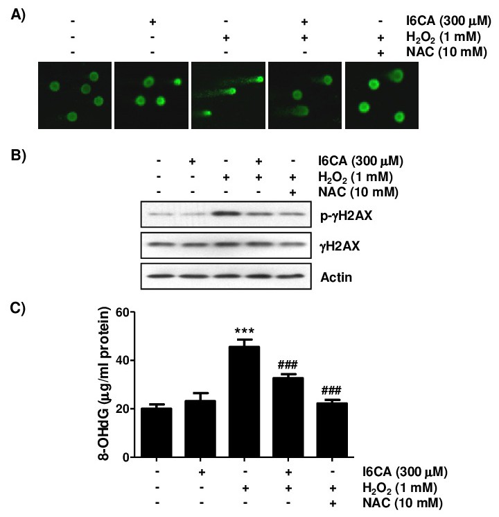
Fig. 4. Protection of H2O2-induced DNA damage by I6CA in V79-4 cells. The cells were treated with or without 300 μM I6CA or 10 mM NAC for 1 h before treatment with 1 mM H2O2 for 24 h. (A) A comet assay was performed, and representative images were captured using a fluorescence microscope (original magnification, ×200). (B) The cell lysates were prepared, and p-gH2AX and gH2AX expression was identified by Western blot analysis. The equivalent loading of proteins in each well was confirmed by actin. (C) The DNA samples of cells were subjected to assessment of the 8-OHdG levels. The measurements were made in triplicate, and the results are expressed as the mean ± SD (***p<0.001 compared with the control group; ###p<0.001 compared with the H2O2-treated group).